The human eye operates as a sophisticated optical system where every component plays a crucial role in vision. Among these components, the eye lens stands out as a remarkable structure that works in harmony with surrounding chambers to focus light precisely onto the retina. Understanding how the eye lens is positioned within the eye’s internal architecture reveals fascinating insights into the mechanics of human vision. The posterior chamber, a fluid-filled space behind the iris, serves as the critical environment where the eye lens maintains its position and function. This intricate arrangement allows light to pass through multiple refractive surfaces before reaching the retina, enabling clear vision at various distances. Exploring the relationship between the posterior chamber and the lens helps us appreciate the complexity of ocular anatomy and understand why certain eye conditions develop when this delicate balance is disrupted.
Table of Contents
ToggleInternal Structure of the Eye: The Relationship Between the Pupil and the Posterior Chamber
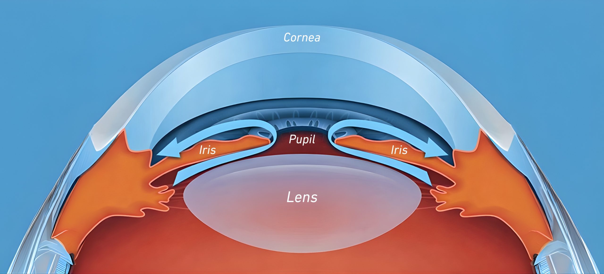
The eye’s internal architecture consists of interconnected compartments that work together to maintain optical clarity and proper pressure balance. The anterior segment of the eye, located in front of the lens, divides into two distinct chambers separated by the iris. The pupil, which appears as the dark circular opening in the center of the iris, serves as the gateway through which light enters the deeper structures of the eye. Behind this adjustable aperture lies the posterior chamber, a narrow space that plays a vital role in nourishing the eye lens and maintaining intraocular pressure.
- Anterior Chamber: This space extends from the cornea at the front of the eye to the iris. It contains aqueous humor, a clear fluid that provides nutrients to avascular structures like the cornea and lens while helping maintain the eye’s shape and pressure.
- Iris and Pupil Connection: The iris functions as a muscular diaphragm that controls pupil size in response to lighting conditions. The pupil itself is not a structure but rather an opening that allows light to pass through to the posterior chamber and lens beyond.
- Posterior Chamber Position: Situated directly behind the iris and in front of the lens, this chamber forms a triangular space in cross-section. It communicates with the anterior chamber through the pupil, allowing aqueous humor to flow forward.
- Ciliary Body Boundary: The posterior chamber is bounded laterally by the ciliary body, which produces aqueous humor and contains the ciliary muscles responsible for accommodation. This structure connects to the lens through delicate zonular fibers.
- Fluid Circulation Pathway: Aqueous humor produced by the ciliary body fills the posterior chamber before flowing through the pupil into the anterior chamber, creating a continuous circulation system that maintains ocular health and pressure.
What is the Posterior Chamber? Its Location and Function in the Eye
The posterior chamber represents a specialized anatomical space within the eye that serves multiple critical functions beyond simply existing as an empty cavity. This chamber occupies the region between the posterior surface of the iris anteriorly and the anterior surface of the lens and ciliary zonules posteriorly. Unlike the larger vitreous cavity that fills most of the eye’s volume behind the lens, the posterior chamber is relatively small but essential for maintaining the health and function of surrounding structures.
The primary function of the posterior chamber involves housing and circulating aqueous humor, the clear fluid that nourishes the avascular eye lens and other structures lacking direct blood supply. The ciliary body, which forms the lateral boundary of this chamber, actively secretes aqueous humor into this space. This fluid then flows forward through the pupil into the anterior chamber, where it eventually drains through the trabecular meshwork at the angle formed by the iris and cornea. This continuous circulation ensures that metabolic waste products are removed while fresh nutrients reach the lens and cornea.
Beyond fluid circulation, the posterior chamber provides the anatomical space necessary for the zonular fibers that suspend the eye lens in its proper position. These delicate fibers extend from the ciliary body to the lens equator, passing through the posterior chamber to maintain lens stability. The chamber also plays a role in the accommodation process, as changes in ciliary muscle tension affect the zonular fibers within this space, allowing the lens to change shape for focusing at different distances. Understanding the posterior chamber’s location and function helps explain why disruptions in this area can lead to significant vision problems, including glaucoma when fluid drainage becomes impaired or lens dislocation when zonular support fails.
How is the Eye Lens Positioned in the Posterior Chamber?
The eye lens maintains its precise position through an elegant suspension system that allows both stability and flexibility. This positioning mechanism involves multiple anatomical components working in coordination to hold the lens in the optical axis of the eye while permitting the shape changes necessary for accommodation. Understanding this positioning system reveals how the eye achieves clear focus across varying distances.
- Zonular Fiber Attachment: The lens is suspended by numerous delicate zonular fibers, also called suspensory ligaments or zonules of Zinn. These transparent fibers extend from the ciliary body and attach to the lens equator, creating a radial support system that holds the lens in position behind the iris and pupil.
- Ciliary Body Anchoring: The zonular fibers originate from the ciliary body, a ring-shaped structure that encircles the lens. This body contains both the ciliary epithelium that produces aqueous humor and the ciliary muscles that control lens shape. The fibers emerge from the valleys and peaks of the ciliary processes, distributing tension evenly around the lens circumference.
- Capsular Insertion Points: The zonular fibers insert into the lens capsule, a transparent elastic membrane that completely envelops the lens substance. These insertions occur primarily at the lens equator, though some fibers attach slightly anterior and posterior to this region, creating a complex network that distributes mechanical forces.
- Tension Balance System: When the ciliary muscles relax, the zonular fibers pull the lens capsule taut, flattening the lens for distance vision. Conversely, when the ciliary muscles contract, tension on the zonules decreases, allowing the elastic lens capsule to mold the lens into a more rounded shape for near vision.
- Posterior Chamber Integration: The entire suspension system operates within and around the posterior chamber. The chamber provides the necessary space for zonular fiber movement during accommodation while the aqueous humor within it bathes the lens surfaces, providing nutrients and maintaining the proper refractive environment.
- Optical Axis Alignment: The zonular support system positions the eye lens precisely along the eye’s optical axis, ensuring that light passing through the pupil encounters the lens center. This alignment is crucial for minimizing optical aberrations and achieving sharp retinal images.
The Relationship Between Posterior Chamber Fluids and Intraocular Pressure
The aqueous humor filling the posterior chamber plays a fundamental role in maintaining intraocular pressure, which is essential for preserving the eye’s shape and ensuring optimal optical function. This clear fluid, continuously produced by the ciliary epithelium, enters the posterior chamber and creates a dynamic pressure system that affects every structure in the anterior segment of the eye. The balance between aqueous production and drainage determines whether intraocular pressure remains within healthy ranges or becomes elevated, potentially leading to vision-threatening conditions.
Aqueous humor production occurs through both active secretion and passive diffusion processes in the ciliary body. Once secreted into the posterior chamber, the fluid flows around the eye lens and through the pupil into the anterior chamber. This forward flow creates a gentle pressure gradient that helps maintain the position of the iris and lens while providing nutrients to these avascular structures. The fluid then exits the eye primarily through the trabecular meshwork, a spongy tissue located at the angle where the iris meets the cornea. A smaller portion drains through the uveoscleral pathway, passing through the ciliary muscle and into the surrounding tissue spaces.
When the delicate balance between aqueous production and drainage becomes disrupted, intraocular pressure can rise to dangerous levels. Blockage of the drainage pathways, whether due to structural abnormalities, inflammation, or other factors, causes fluid to accumulate in both the posterior and anterior chambers. This pressure increase can compress the optic nerve at the back of the eye, leading to glaucoma and progressive vision loss if left untreated. Conversely, decreased aqueous production or excessive drainage can result in abnormally low intraocular pressure, potentially causing the eye to lose its proper shape and compromising vision. The posterior chamber’s role in this pressure system makes it a critical focus area for understanding and treating pressure-related eye diseases. Monitoring and managing intraocular pressure through regular eye examinations helps preserve the health of the eye lens, optic nerve, and other vital structures.
Posterior Chamber Problems: Connection to Eye Diseases
Disruptions in the posterior chamber’s normal anatomy and function can lead to various eye diseases that significantly impact vision and ocular health. Understanding these conditions helps explain why maintaining the integrity of this small but crucial space is essential for preserving sight. The posterior chamber’s involvement in fluid dynamics, lens support, and pressure regulation means that problems in this area often have widespread effects throughout the eye.
Angle-closure glaucoma represents a serious condition where the iris moves forward and blocks the drainage angle, preventing aqueous humor from flowing properly from the posterior chamber through the pupil. This blockage causes rapid pressure buildup that can damage the optic nerve within hours if not treated promptly. The condition may occur acutely, causing severe eye pain, blurred vision, and nausea, or develop gradually in a chronic form. Anatomical factors such as a shallow anterior chamber, thick lens, or narrow angle increase the risk of this condition, particularly in farsighted individuals and older adults whose lenses naturally thicken with age.
Pigment dispersion syndrome occurs when pigment granules from the posterior surface of the iris break free and circulate through the posterior chamber and anterior chamber. These particles can clog the trabecular meshwork, impairing aqueous drainage and leading to elevated intraocular pressure. Young, nearsighted males are particularly susceptible to this condition, which may progress to pigmentary glaucoma if the drainage system becomes significantly compromised. The mechanical rubbing between the iris and zonular fibers in the posterior chamber contributes to pigment release, especially during activities that cause pupil dilation and constriction.
Lens subluxation or dislocation involves displacement of the eye lens from its normal position due to weakened or broken zonular fibers. Trauma, genetic connective tissue disorders, or age-related zonular weakness can cause the lens to shift partially or completely out of position. When the lens moves into the posterior chamber or vitreous cavity, it can block aqueous flow and cause acute pressure elevation. Even partial lens displacement affects vision quality and may require surgical intervention to reposition or remove the unstable lens. The posterior chamber’s role as the site of zonular attachment makes it central to understanding and treating lens displacement disorders.
Conditions Requiring Intervention in the Posterior Chamber and Lens
Several medical and surgical interventions target the posterior chamber and eye lens when disease or age-related changes compromise vision or threaten eye health. Cataract surgery represents the most common procedure involving this anatomical region, performed when the natural lens becomes cloudy and impairs vision. During this procedure, surgeons access the lens through the anterior chamber, carefully remove the clouded lens material while preserving the lens capsule, and implant an artificial intraocular lens in the same position. The posterior chamber’s anatomy remains largely intact during modern cataract surgery, with the new lens positioned where the natural lens once sat, supported by the remaining capsule and zonular structures.
Laser peripheral iridotomy provides treatment for angle-closure glaucoma or narrow angles at risk of closure. This procedure creates a small opening in the peripheral iris, allowing aqueous humor to flow directly from the posterior chamber to the anterior chamber even if the pupil becomes blocked. By equalizing pressure between the two chambers, the laser treatment prevents the iris from bowing forward and blocking the drainage angle. This relatively simple outpatient procedure can prevent vision-threatening acute angle-closure attacks in susceptible individuals, preserving the normal function of the posterior chamber and protecting the optic nerve from pressure damage.
Surgical management of lens subluxation or dislocation may become necessary when zonular support fails and the eye lens shifts from its proper position in the posterior chamber. Depending on the extent of zonular damage and lens displacement, surgeons may employ various techniques to stabilize or replace the lens. In cases of partial zonular loss, capsular tension rings or segments can be implanted to support the remaining zonules and maintain lens position. When zonular support is severely compromised, surgeons may remove the unstable natural lens and secure an artificial lens using alternative fixation methods, such as suturing to the iris or sclera, or placing the lens in the anterior chamber. These interventions restore clear vision while preventing complications like glaucoma or retinal detachment that can occur when a displaced lens disrupts normal posterior chamber function.
Why is Understanding Eye Anatomy Important? Health Awareness and Preventive Care
Knowledge of eye anatomy, particularly the relationship between the posterior chamber and eye lens, empowers individuals to recognize symptoms that warrant professional evaluation and understand the rationale behind recommended treatments. When people comprehend how their eyes function and what can go wrong, they become more motivated to maintain regular eye examinations and adopt protective behaviors. The eye’s complexity means that subtle changes in structures like the posterior chamber can have significant consequences for vision, making early detection through routine screening essential for preserving sight throughout life.
Understanding the posterior chamber’s role in maintaining intraocular pressure helps explain why eye care professionals measure pressure during comprehensive examinations. Elevated pressure often produces no noticeable symptoms until significant optic nerve damage has occurred, making glaucoma a silent threat to vision. By knowing that the posterior chamber participates in the fluid dynamics that determine eye pressure, patients better appreciate why pressure monitoring and treatment compliance are crucial for preventing irreversible vision loss. This knowledge also helps individuals recognize risk factors such as family history, age, and certain anatomical features that increase glaucoma susceptibility, prompting more vigilant monitoring.
Awareness of how the eye lens is positioned and supported within the posterior chamber provides context for understanding age-related vision changes and the need for cataract evaluation. As people age, the lens naturally becomes less flexible and may develop clouding that gradually impairs vision. Knowing that the lens can be safely removed and replaced with an artificial version helps reduce anxiety about cataract surgery, one of the most successful and commonly performed procedures in medicine. Additionally, understanding zonular fiber function explains why certain genetic conditions, injuries, or inflammatory diseases can affect lens position, highlighting the importance of comprehensive eye examinations that assess not just visual acuity but also anatomical integrity. This anatomical knowledge transforms patients from passive recipients of care into informed participants who can make educated decisions about their eye health and recognize when professional intervention is needed to preserve their precious gift of sight.